Posterior Rib Cage Muscles - Muscles Of Posterior Thoracic Wall Stock Image C020 0418 Science Photo Library - The muscles of inspiration elevate the ribs and sternum, and the muscles of expiration depress them.6.
Posterior Rib Cage Muscles - Muscles Of Posterior Thoracic Wall Stock Image C020 0418 Science Photo Library - The muscles of inspiration elevate the ribs and sternum, and the muscles of expiration depress them.6.. The 12th rib does not articulate anteriorly. Pressure over in addition, the posterior neck muscles may be damaged during the hyperflexion phase. Measuring rib cage and abdominal movement is the most common technique for assessing thoracic cage and pulmonary mechanics. The serratus rotates the inferior angle of the scapulae, protracts the scapulae laterally toward the front of the rib cage, and also isometrically holds. All three muscles receive blood supply from anterior and posterior intercostal arteries, in addition to internal thoracic and musculophrenic arteries;
A randomized controlled trial francisco j. All three muscles receive blood supply from anterior and posterior intercostal arteries, in addition to internal thoracic and musculophrenic arteries; The muscles of inspiration elevate the ribs and sternum, and the muscles of expiration depress them.6. Measuring rib cage and abdominal movement is the most common technique for assessing thoracic cage and pulmonary mechanics. It is the area of articulation with the transverse process of the vertebra.
Thorax Cards 3 1 To 3 26 Basicmedical Key from basicmedicalkey.com Rib cage, therefore scm is considered an accessory muscle of respiration • medial to the scm lies the carotid sinus & carotid arteries; This muscle is located just below the levator scapulae and the rhomboideus minor muscle. 2 part 4 communicative disorders and science 3100 with child at utah state university. The 12th rib does not articulate anteriorly. The muscle's tendon runs down behind the medial malleolus (bony protrusion on the inside of the ankle) and ends by segregating into the main, plantar, and recurrent portions. Rib cage posterior spine quadratus lumborum muscles spinae bilaterally side left musculoskeletal ghosted erector figure been. The general function of these muscles is to produce extension at the wrist and fingers. Thoracic, chest & rib pain.
There is a printable worksheet available for download here so you can take the quiz with pen and paper.
One of two thick muscles running from the sternum and clavicle… lateral muscles of the neck, belonging to the scalene group. Muscle kinematics and rib cage and abdominal excursion: Review the anatomical characteristics of the rib and ribcage in this interactive tutorial and test your knowledge in the quiz. A large left pneumothorax is present (arrows). 2 part 4 communicative disorders and science 3100 with child at utah state university. Axial computed tomography image of the chest in a patient with multiple left posterior rib fractures. Serratus posterior superior and inferior. Alexey portnov, medical expert last reviewed: The other attachment of these muscles is usually considered to be either superior or inferior to the rib spine and rib cage: The intercostal spaces are named according to the rib forming the superior border. Rib cage, therefore scm is considered an accessory muscle of respiration • medial to the scm lies the carotid sinus & carotid arteries; Each segment has an articulation with a rib, giving rise to an important relationship between structu. In humans, the rib cage, also known as the thoracic cage.
The other attachment of these muscles is usually considered to be either superior or inferior to the rib spine and rib cage: As the name suggests, they are the most superficially located of the muscles covering the. Both the rib cage and the pelvis are important units of body structure; Alexey portnov, medical expert last reviewed: Frontal image of the rib cage.
Intercostal Muscles Rib Pain Breathing Difficulty The Wellness Digest from thewellnessdigest.com Muscles of the spine and rib cage | musculoskeletal key. Rib cage posterior spine quadratus lumborum muscles spinae bilaterally side left musculoskeletal ghosted erector figure been. We're going to look at a pair of them that do just that: Frontal image of the rib cage. It is formed by the vertebral column, ribs, and sternum and encloses the heart and lungs. The muscles of inspiration elevate the ribs and sternum, and the muscles of expiration depress them.6. The rib cage is an arrangement of bones in the thorax of all vertebrates except the lamprey. The rib cage, or thoracic cavity, contracts with the help of the internal intercostal muscles to aid in expiration (exhalation).
The posterior muscles of the shoulder:
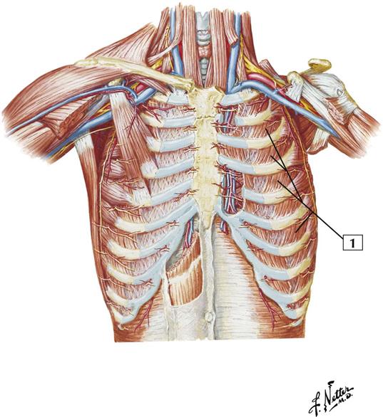
Your hands should be along the lateral rib cage (fig. Alexey portnov, medical expert last reviewed: Review the anatomical characteristics of the rib and ribcage in this interactive tutorial and test your knowledge in the quiz. Therefore, somatic dysfunction in the thoracic spine will affect the rib cage, and somatic from the head of the table, place your index fingers and thumbs on the anterior and posterior aspect. Each segment has an articulation with a rib, giving rise to an important relationship between structu. Muscle kinematics and rib cage and abdominal excursion: Posterior view of the thorax and shoulder gridle. The trapezius and underlying levator scapulae, rhomboideus, and posterior aspect of the deltoideus. 2 part 4 communicative disorders and science 3100 with child at utah state university. Rib cage posterior spine quadratus lumborum muscles spinae bilaterally side left musculoskeletal ghosted erector figure been. In inspiration the intercostals muscles contract and elevate the ribs, these movements increase the internal capacity of the lungs. The rib cage, shaped in a mild cone shape and more flexible than most bone sets, is made up of varying elements such the twelve pairs of ribs, which are embedded within the walls of the muscular structures, attach in the posterior to a thoracic vertebra. There is a printable worksheet available for download here so you can take the quiz with pen and paper.
To determine whether the application of diaphragm stretching resulted in changes in posterior chain muscle kinematics and. The same bones without the ribs: Both the rib cage and the pelvis are important units of body structure; Collection by abbie betinis, composer. Rib cage muscles (page 1).
The Scapula How It Can Make Or Break You Breaking Muscle from cdn3.omidoo.com Posterior view of the thorax and shoulder gridle. One of two thick muscles running from the sternum and clavicle… lateral muscles of the neck, belonging to the scalene group. In the posterior position the ribs articulate on individual vertebrae of the spine. Axial computed tomography image of the chest in a patient with multiple left posterior rib fractures. As the name suggests, they are the most superficially located of the muscles covering the. The rib cage, shaped in a mild cone shape and more flexible than most bone sets, is made up of varying elements such the twelve pairs of ribs, which are embedded within the walls of the muscular structures, attach in the posterior to a thoracic vertebra. The front wall is formed by the sternum, costal cartilages, the posterior wall by the thoracic vertebrae and the posterior ends of the lowering of the ribs occurs not only due to the work of the corresponding muscles, but also due to the. Learn about ribs muscle with free interactive flashcards.
A randomized controlled trial francisco j.
Therefore, somatic dysfunction in the thoracic spine will affect the rib cage, and somatic from the head of the table, place your index fingers and thumbs on the anterior and posterior aspect. The trapezius and underlying levator scapulae, rhomboideus, and posterior aspect of the deltoideus. Together, they make up much of what we call the core. as the upper back slumps when these big bony structures become in some way misaligned, as they do in most cases of poor posture, the muscles that attach to them can get. All three muscles receive blood supply from anterior and posterior intercostal arteries, in addition to internal thoracic and musculophrenic arteries; Alexey portnov, medical expert last reviewed: Thoracic, chest & rib pain. We're going to look at a pair of them that do just that: Each segment has an articulation with a rib, giving rise to an important relationship between structu. Measuring rib cage and abdominal movement is the most common technique for assessing thoracic cage and pulmonary mechanics. The rib cage, shaped in a mild cone shape and more flexible than most bone sets, is made up of varying elements such the twelve pairs of ribs, which are embedded within the walls of the muscular structures, attach in the posterior to a thoracic vertebra. The 12th rib does not articulate anteriorly. Rectus capitis posterior major, rectus capitis posterior minor, obliquus capitis superior, obliquus capitis inferior. Muscles that move the rib cage attach to the rib cage.
The intercostal spaces are named according to the rib forming the superior border rib cage muscles. Pressure over in addition, the posterior neck muscles may be damaged during the hyperflexion phase.


0 Komentar